
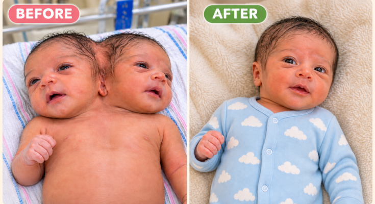
A newborn diagnosed with the rare condition known as dicephalic parapagus — two fully formed heads and brains sharing a single torso and lower body — underwent a lengthy, high-risk supportive surgical procedure late Thursday at St. Brigid’s Children’s Hospital. Hospital officials confirmed Friday that the infant came through the operation and is currently stable in the pediatric intensive care unit, where continuous monitoring continues.
The case has drawn quiet attention within medical circles and, after a photograph circulated online earlier in the week, among the wider public. Hospital staff and the family have asked for privacy as treatment proceeds. What follows is an account compiled from statements by the medical team, a brief family statement released through the hospital, and background on the condition itself.
The Birth and Initial Diagnosis
The infant was born earlier in the week. According to hospital officials, the delivery itself was managed by a multidisciplinary team that had been on standby after prenatal imaging raised the possibility of conjoined twinning. Dicephalic parapagus is a subtype of parapagus twinning in which two heads sit side by side on one body. Each head typically has its own brain and spinal cord extending into the upper chest; the degree of sharing of the heart, lungs, and abdominal organs varies from case to case.
In this instance, initial postnatal scans and clinical examination showed two distinct heads, two brains, and two hearts, with the majority of the abdominal organs and the entire lower body shared. Dr. Anika Rao, the pediatric surgeon leading the care team, described the anatomy in a briefing held before the operation as “a classic presentation.” She noted that full separation into two independent survivors is not considered possible. “The shared structures below the upper chest make it impossible to divide the body in a way that would allow both to live separately,” she said. “Our efforts from the beginning have been directed toward supporting the shared physiology and addressing any complications that arise.”
The parents, Elena and Marcus Vale, have remained largely out of public view. In the short statement they authorized the hospital to release before surgery, they said only that they were concentrating on the immediate needs of their children and placing their trust in the medical team. Hospital staff described them as present at the bedside whenever visiting rules allowed, one parent often resting a hand near each head.
Preoperative Condition and Planning
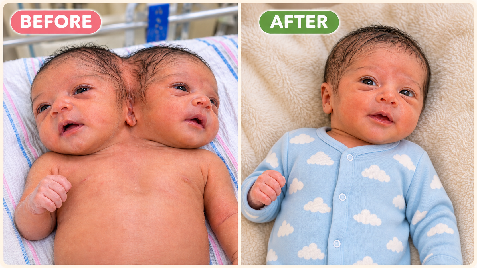
In the days after birth the infants were stable enough to begin feeding, though both required close observation. One heart rate ran consistently a little faster than the other — a detail the team noted but did not treat as an emergency on its own. Respiratory effort, however, became more labored as the week progressed. Imaging and blood-flow studies suggested that the shared vascular pathways and the configuration of the upper airways were creating inefficient circulation and intermittent difficulty with oxygenation.
Dr. Rao explained that the surgical plan was never intended as an attempt at separation. Instead, the team prepared a targeted intervention lasting many hours: adjustments to the shared vascular connections to improve balance between the two hearts, and procedures to secure better airway access so that ventilatory support could be delivered more effectively. “These are supportive measures,” she said. “They do not change the fundamental anatomy. They are meant to give the shared body a better chance to function while we continue to learn what each infant needs.”
A large operating-room team was assembled, including pediatric cardiac and general surgeons, neonatologists, anesthesiologists experienced in complex congenital cases, and specialized nursing staff. The hospital’s ethics committee had reviewed the plan earlier in the week. No public details of that review were released, but officials confirmed that the discussion centered on the balance between potential benefit and the significant risks inherent in operating on such a fragile, shared physiology.
The Operation
The procedure began late Thursday and continued for more than seven hours. Hospital spokespeople provided only limited updates during the night, stating at intervals that the operation was proceeding as planned and that no major unexpected events had been reported from the operating room. By early Friday morning the team had finished the planned work.
In a brief postoperative statement, Dr. Rao said the vascular adjustments and airway procedures had been completed successfully. “Both hearts continued to function throughout, and we were able to improve the balance of blood flow,” she reported. “The infants are receiving the ventilatory support we anticipated after a surgery of this length and complexity. We have not identified any new complications beyond those we prepared for.”
Outside experts, speaking in general terms about dicephalic parapagus rather than this specific case, noted that any operation on shared thoracic and upper-abdominal structures carries substantial risk. They also observed that supportive procedures of this kind, while sometimes able to stabilize immediate problems, do not alter the long-term reality of a single body supporting two nervous systems and two hearts.
Immediate Recovery
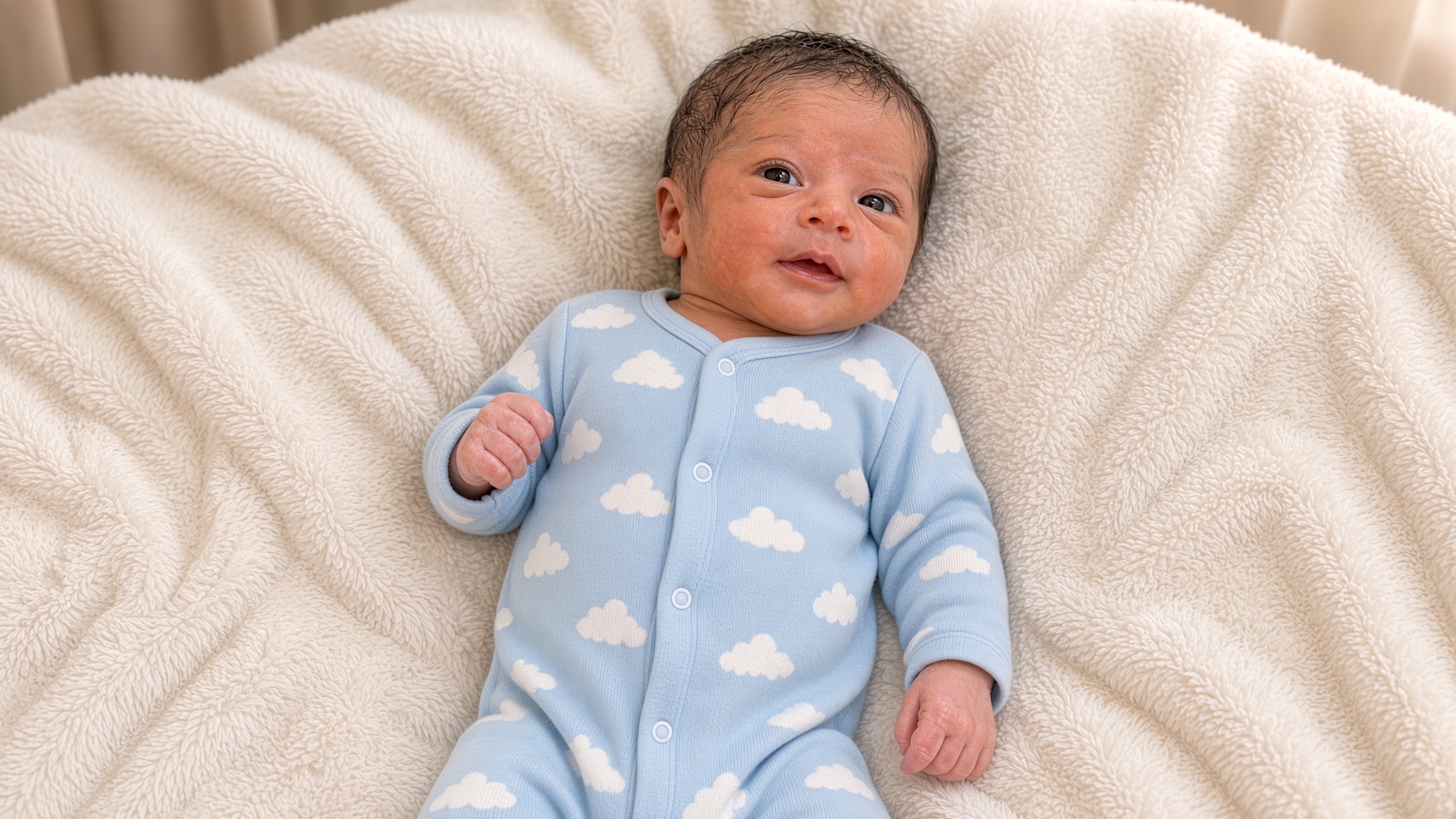 As of Friday afternoon the infant remained in the pediatric intensive care unit under continuous monitoring. Vital signs were described as stable within the parameters expected after major surgery. Both heads showed periods of spontaneous movement and response to light touch, though the effects of anesthesia and pain medication made detailed neurological assessment premature. Feeding has not yet been restarted; nutrition is being provided intravenously for the time being.
The Vales have been permitted short visits. Hospital staff said the parents sit quietly beside the warmer, speaking softly and occasionally resting a hand on an arm or shoulder. No further public statement has been issued by the family.
Background on the Condition
Dicephalic parapagus is one of the less common forms of conjoined twinning. Overall, conjoined twins occur in roughly one in 50,000 to 200,000 births, with a higher proportion of female infants. Among the various types — thoracopagus (joined at the chest), omphalopagus (joined at the abdomen), pygopagus (joined at the lower back), ischiopagus (joined at the pelvis), and craniopagus (joined at the skull) — parapagus forms involve side-by-side fusion and frequently include the dicephalic presentation of two heads on one body.
Medical literature records only a small number of individuals with dicephalic parapagus anatomy who have survived into childhood or adulthood. The best-known living examples are Abigail and Brittany Hensel of Minnesota, each of whom has her own brain, heart, and spinal cord while sharing the lower body; they have coordinated the shared limbs well enough to walk, attend school, drive, and work as teachers. Such outcomes are exceptional and require years of highly individualized medical, physical, and occupational support. Most pregnancies involving conjoined twins do not result in live birth, and among those that do, survival beyond the first days or weeks remains uncommon when major organs are shared.
In the present case, hospital officials have declined to offer any prognosis beyond the immediate postoperative period. “Every anatomy is different,” Dr. Rao said. “We are taking each day as it comes and focusing on comfort, stability, and clear communication with the family.”
Hospital Response and Public Attention
St. Brigid’s has not released photographs or additional clinical images. Officials confirmed that the picture which circulated online earlier in the week depicts the infant, but they have asked media outlets and the public not to redistribute it out of respect for the family’s privacy. The hospital’s communications office has limited its comments to the factual updates summarized above.
Inside the unit, staff describe a deliberately quiet atmosphere. Extra nursing coverage has been assigned, and the room is arranged so that monitors and equipment do not crowd the warmer more than necessary. A social worker and a child-life specialist have met with the parents to discuss practical support and longer-term resources, including connections to the small number of centers that have previously cared for dicephalic parapagus patients.
Looking Ahead
Further imaging is scheduled over the coming days to evaluate the results of the vascular and airway work. The medical team will also watch for signs of infection, bleeding, or changes in heart or lung function — all standard concerns after prolonged surgery in a neonate. Decisions about when and how to resume feeding, and about any additional supportive measures, will be made day by day.
For the Vales, the immediate horizon remains the next set of vital-sign checks and the next brief visit. In their earlier statement they asked only for space to focus on their children. Hospital staff say that request continues to guide the care being provided.
As of the latest update Friday evening, the infant’s condition was listed as stable. Both hearts were beating, the shared body was receiving mechanical ventilatory support, and the parents remained at the bedside. No further surgery is planned at this time. The medical team, the family, and the hospital have indicated that any additional information will be released only when there is a material change in condition or when the parents choose to speak further.
